As we have seen in successive articles, fetal growth is a complex multifactorial phenomenon that depends on genetic and environmental factors. In some cases intrauterine growth retardation may occur (RCIU), when there is a mismatch that prevents the proper development of the fetus.
It refers to the poor growth of a baby while in the womb during pregnancy, specifically, that the fetus weighs less than 90% than other babies of the same gestational age.
This phenomenon is also called "restricted intrauterine growth" to define a baby that is smaller than normal during pregnancy: babies do not grow inside the uterus at the speed they should and usually have a lower birth weight.
The most common cause of fetus growth problems is in a malfunction of the placenta, which is the tissue that carries food and oxygen to the baby. Although as we have seen on multiple occasions, there are other factors that influence fetal growth, such as genetic alterations, malformations, consumption of tobacco or drugs and high blood pressure before or during pregnancy ...
X-ray exposures and infections during pregnancy that affect the fetus, such as rubella, cytomegalovirus, toxoplasmosis and syphilis, can also affect fetal weight.
As we can see, there are some causes on which we cannot act (or it will be done medically), but others respond to the mother's health habits (or rather poor health) and are controllable, such as tobacco, alcohol or other consumption. type of drugs that could cause the baby not to grow properly inside the uterus.
Types of fetal growth retardation
Depending on the cause of this delay, the fetus may be symmetrically small or have a head of normal size for its gestational age, while the rest of its body is small. In this sense, three types of RCIU are described, based on the incorporation into the clinic of the concept of the three phases of cell growth described by Winnick:
RCIU type I or symmetric, occurs when in the phase of cellular hyperplasia (which occurs in the first 16 weeks of fetal life) damage occurs with a decrease in the total number of cells. In these newborns there is a symmetrical growth of the head, abdomen and long bones.
RCIU type II or asymmetric, occurs when in the phase of cellular hypertrophy, which occurs after 32 weeks of gestation and lasts approximately 8 weeks. It is characterized by a disproportionate growth between the head and long bones and the fetal abdomen.
RCIU type III or mixed, occurs between 17 and 32 weeks gestation, in the phase of hyperplasia and concomitant hypertrophy and the appearance will depend on the time in which the injury occurs.
Another classification is based on the etiology or origin of the disorder:
- Intrinsic RCIU, mainly for causes that are in the same fetus, such as chromosomal defects.
- Extrinsic RCIU, in this case the causes are external elements to the fetus, such as a placental pathology.
- Combined RCIU, in which a combination of the above factors are presented.
- Idiopathic RCIU, in which the cause of the fetus growth disorder is unknown.

How fetal growth retardation is detected
Intrauterine growth retardation It can be suspected if the size of the uterus of the pregnant woman is small. The condition is usually confirmed by ultrasound. The measurement from the mother's pubic bone to the upper part of the uterus will be smaller than expected for the gestational age of her baby. This measurement is called the height of the uterine fundus.
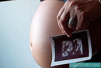
During pregnancy throughout the various controls the doctor can determine if the baby is growing normally. The main test to monitor the growth of a baby in the womb is ultrasound, which allows you to take a series of measurements of the baby to assess the weight.
In addition, ultrasound allows studying the functioning of the placenta through a technique called Doppler, which currently controls hypertension or diabetes in the mother, cardiac malformations and problems with the umbilical cord and placenta, the main factors that can endanger the health of the unborn baby.
Ultrasound also allows you to determine the amount of amniotic fluid and the movements that the baby performs because some babies with intrauterine growth retardation have a decrease in the amount of amniotic fluid and movements. If the baby is detected to be small, ultrasound scans are performed more frequently.
Additional tests may be needed to detect infection or genetic problems if such intrauterine growth retardation is suspected, since the RCIU increases the risk of the baby dying inside the uterus before birth. If the doctor suspects that the mother could have this condition, she will be carefully monitored with some ultrasound of the pregnancy to measure the growth, movements, circulation and fluid around the baby.
Although we must point out that not all small babies have stunting in the womb. Only one third of babies who are small at birth have stunted growth. The rest are simply smaller than normal. Like adults, there are children of different sizes and genetics at this point has enough to say for the baby to be more or less large, without having a developmental delay.


